
Our Aching Joints
Many Causes, Many Treatments

UPDATED: August 3, 2026
Welcome to the Healthy Living Is Good Medicine Newsletter, presenting timely, science-based, original articles covering a wide range of preventive medicine and public health topics, along with critical commentaries on the politics and economics of the American healthcare system.
Musculoskeletal Injuries and Chronic Pain
I’m not a specialist in orthopedic medicine or surgery, and I’m not an expert in joint problems. My experience with painful joints primarily comes from a decade spent as the medical administrator of a multi-disciplinary clinic devoted to the rehabilitation of musculo-skeletal injuries involving the spine and extremities. Our clinic offered individualized integrated programs, and provided a medical alternative to chiropractic care for back and neck pain.
We employed a conservative (non-invasive) approach to the management of chronic pain and associated disabilities. From a “quality control” perspective, I monitored what was and wasn’t working, and made sure that patients were quickly referred to appropriate outside specialists when they didn’t make satisfactory progress with our programs.
It was very satisfying to see that most of our patients were able to return to full, pain-free functioning, without the need for operative interventions. However, when the non-surgical options proved ineffective, and surgery was clearly the treatment of choice, my job was to encourage patients to seek treatment with an appropriate surgical specialist. As a result, I became very familiar with patient resistance to surgery.
I subsequently experienced that same kind of resistance in myself, following a major rotator cuff injury. Expecting tendon reattachment surgery when I saw a highly respected orthopedic surgeon assigned to the U.S. Ski Team, she insisted that shoulder joint replacement was my only choice if I wanted to be able to use my arm to its full extent, pain-free. So, I got a second opinion.
I was really bummed out when the second surgeon agreed with the first one. I simply didn’t want to undergo the long and daunting post-op rehab that would be required for a reverse total shoulder arthroplasty (RTSA), so I insisted on a prescription for physical therapy. Fortunately, my shoulder rehab program, crafted by a couple of very experienced physical therapists, proved both surgeons wrong.
To be clear, my ruptured tendons did not mend, and my shoulder will never be as good as it once was, but it works well enough for all my activities of daily living, and then some. This anecdote may or may not be applicable to others with a similar injury. As they say, “your mileage may vary.” Just be sure that when considering non-surgical options, you don’t fall prey to scammers.
Under America’s for-profit healthcare system, orthopedic surgeons tend to view joint problems from the perspective of expensive surgical solutions. In countries with a taxpayer funded national health service, cost containment is a priority and preventive measures receive the emphasis they deserve. Keep in mind that there may be other acceptable, non-surgical options available, but patients may have to take the initiative to explore them.
In marked contrast to seeking conservative care was my personal experience with a herniated lumbar disc. Accompanied by spinal stenosis, there was no reasonable alternative to surgery, and delaying it could result in persistent neurological deficits. I was able to find a neurosurgeon who specialized in micro-discectomy procedures. I underwent outpatient surgery and was discharged pain-free, with normal nerve function and a tiny incision.
As an aside, although there are orthopedic surgeons who perform spine surgery, if nerves are involved I would always choose a neurosurgical sub-specialist, because they have trained on nerve tissue from the very get-go. Treating the spinal cord and spinal nerves with utmost care and respect is what matters most to me. Bony fixation procedures don’t require the same degree of precision as neurosurgery.
Although there are many causes and contributors to acute and chronic joint pain, today’s article will focus on osteoarthritis of the knee. I will leave discussions of tendinopathies, ligamentous injuries including strains, sprains, and ruptures, meniscus tears, patellar fractures, and bursitis to someone else. The spinal facet joints deserve an entire article of their own.
If you don’t have aching joints right now, just live long enough, and/or play hard enough, and one day you will. Best be prepared.
Osteoarthritis
The term “arthritis” refers to joint inflammation. Causes can include age-related deterioration of the bone and cartilage (primary osteoarthritis), and secondary causes such as traumatic injuries to joint structures, congenital deformities, infection (septic arthritis), metabolic dysfunction causing deposition of uric acid (gout), or an auto-immune disease such as psoriatic arthritis, rheumatoid arthritis, or lupus.
Osteoarthritis (OA) is the most common form of arthritis, usually as primary osteoarthritis. Its presentation varies widely in severity of pain and disability. There is a substantial genetic component that’s polygenic and site specific. Twin and family studies indicate genetic susceptibility is a risk factor in roughly 60 to 65 percent for hand OA cases that involve Heberden’s or Bouchard’s nodes. The heritability for hip OA is around 60 percent, and 40 to 50 percent for knees. There are stronger associations in women for hand and knee OA than with men.
Primary osteoarthritis is considered an age-dependent degenerative joint disease. Structural changes that occur in cartilage and bone may eventually cause pain, stiffness, swelling, and decreased joint mobility. Medications can be used to ameliorate inflammation and pain, and knee braces can redistribute loading on the affected joints.
A 2022 review covers the different trauma-related risk factors for developing secondary knee OA. One of the most effective ways to lower the risk of osteoarthritis of the hip, knee, and ankle is to reduce excessive joint loading by losing weight.
Osteoarthritis can involve any joint, although knee and hip joints are the ones most commonly treated with surgical joint replacement. Interestingly, the X-ray findings do not necessarily correlate with the degree of pain or impaired mobility. I distinctly remember being dumbfounded by a patient who had a normal gait and a hip X-ray showing that the joint was obliterated.
Knee Osteoarthritis
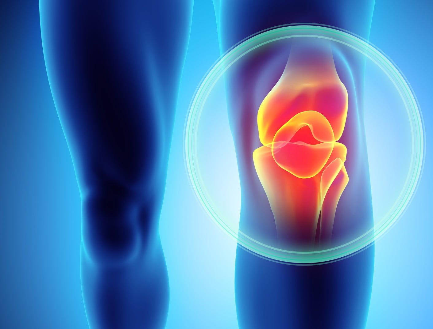
Today’s article will mainly focus on osteoarthritis of the knee because of several subscriber requests. Every year, more than 700,000 Americans undergo knee replacement surgery. Nearly a third of the U.S. population will be affected by osteoarthritis of the knee by age 70.
My main ski buddy has had one knee replaced, and the other one is long overdue. Most recently, a close friend underwent total knee replacement, and her challenging post-operative recovery inspired us to co-create this song:
Degrees of Severity
The standard system for staging knee osteoarthritis on X-ray is the Kellgren-Lawrence (K-L) grading scale:
K-L Grade 0 (Normal): No joint space narrowing, no osteophytes (bone spurs), no sclerosis (scarring).
K-L Grade 1 (Doubtful): This grade indicates uncertainty, rather than definitive disease.
K-L Grade 2 (Mild or Minimal): Bony outgrowths at the joint margins are present. Possible joint space narrowing on the anteroposterior weight-bearing film, indicating cartilage loss due to wear. This is generally considered the radiographic diagnostic threshold.
K-L Grade 3 (Moderate): Multiple, moderate-sized osteophytes. Definite narrowing of the joint space. Some sclerosis of subchondral bone seen as increased bone density beneath the cartilage, due to altered loading forces. Possible deformity of the bone contour, such as a flattening of the femoral condyle.
K-L Grade 4 (Severe): Large osteophytes and marked subchondral sclerosis. Profound joint space narrowing, often described as “bone-on-bone.” There are definite bony contour deformities caused by structural remodeling from long-standing biomechanical dysfunction. Subchondral cysts, appearing as fluid-filled cavities within the bone, may be present.
The evidence for changes in weather influencing joint pain may be a real, physiologically plausible phenomenon for a particular subset of osteoarthritis patients. Humidity, falling barometric pressure, and decreasing temperature are the more consistently implicated variables. However, weather would be a relatively modest modulating effect on an already-multifactorial pain syndrome. Although a 2023 systematic review and meta-analysis of 14 observational studies suggests an association between weather and OA pain, the claims that patients can “predict rain with their knee joints” are not supported by population-level data.
Conservative Management
Non-surgical treatments are typically tailored to the individual patient’s biomechanical profile and lifestyle, integrating rehabilitation with selected adjunctive treatments when appropriate. Intra-articular injectables may provide temporary relief, although effectiveness is primarily short-term, and strength of evidence for their benefits varies greatly.
Doctors who specialize in the conservative management of knee osteoarthritis may offer a variety of medications and procedures, but healthcare consumers should be wary of treatments that are not considered a part of science-based, mainstream medicine. In other words, beware of unproven and quack treatments.

Corticosteroid joint injections are mostly safe and tolerated well, but patients and practitioners should be aware of the small risk of adverse effects, as well as the limited duration of their benefits. Proper patient selection is essential, and the timing of injections should avoid proximity to potential surgical dates. Major society guidelines now explicitly state that intra-articular corticosteroid injection should be avoided for at least three months preceding joint replacement surgery to avoid a significantly higher risk of prosthetic infections, according to a 2022 meta-analysis.
Dealing with chronic joint pain by receiving corticosteroid injections every six months is a common stop-gap measure, but intra-articular steroids can accelerate joint deterioration. A 2017 randomized controlled trial (RCT) of repeated triamcinolone injections every three months for two years showed measurable cartilage volume loss on MRI, and provided no better pain relief, compared to a saline placebo.
Cartilage loss and radiographic joint space narrowing following corticosteroid injections for knee osteoarthritis was confirmed by a 2023 systematic review and meta-analysis. Intra-articular normal saline injections, although associated with improvements in both patient-reported pain and function scores, should only be used as the placebo arm in RCTs.

Platelet-rich plasma (PRP) injections employ a concentrated solution of a patient’s own blood platelet cells to stimulate healing of damaged periarticular tissues such as muscles, tendons, and ligaments. Early studies suggest that PRP joint injections may help treat osteoarthritic pain and stiffness by modulating the joint environment and reducing inflammation.
A 2025 systematic review of six RCTs suggests that PRP may be a viable therapeutic alternative for managing knee OA pain, comparable in efficacy to corticosteroid injections. However, there is not yet convincing evidence that PRP injections support intra-articular cartilage repair. As usual, more robust research would be necessary before that claim could be made.
Prolotherapy, also known as proliferation therapy or regenerative therapy, uses injections of a concentrated dextrose (sugar) solution. It purportedly stimulates growth factor production that could potentially promote healing of injured connective tissues. It has been used to treat pain in the knee, hip, ankle, foot, shoulder, elbow, and finger joints. There is currently insufficient evidence to support the routine use of prolotherapy for degenerative joint diseases such as osteoarthritis.
The routine use of hyaluronic acid injections for osteoarthritis is not recommended, although there are subsets of patients who have failed with other therapies who reportedly had benefits from that treatment. Injections of bone marrow aspirate concentrate, stromal vascular fraction, and mesenchymal stem cell injections, employed in some medical settings outside the mainstream, are also not recommended.
Intra-articular injections of so-called “homeopathic remedies” such as a German solution called “Zeel and Traumeel” do not have FDA approval. Homeopathy as a healing practice has been thoroughly debunked. The combo contains extremely diluted herbal ingredients such as Arnica, Echinacea, and Comfrey root, and minerals.
This is a very expensive placebo injection at best, and a risky invasive procedure at worst. Exposing unsuspecting patients to a possible joint infection while performing a sham treatment seems unscrupulous. Doctors who engage in such practices should be reported to their licensing board.
Motion Is Lotion
There is a real and reasonably well-characterized mechanism through which articular cartilage can be preserved, or at least to slow its degeneration. Because cartilage has no blood supply, the only way chondrocytes (cartilage cells) can receive their necessary nutrition is by synovial joint fluid being pumped throughout the cartilage matrix by cyclical loading and unloading. This is the basis for many of the “exercise is chondroprotective” studies.
Terrestrial and aquatic therapeutic exercises consistently show moderate effects for pain reduction and functional improvements in knee osteoarthritis. These studies are as well-replicated as anything else in the OA literature. Effects of exercise on pain are generally comparable in magnitude to the use of NSAIDs, without the risk profile.
There is no compelling evidence that appropriate-intensity exercises accelerate degenerative joint disease progression. High-intensity loading has equivocal data on OA risk, but moderate recreational activities and structured therapeutic exercises do not show an acceleration of cartilage loss.
A 2025 study examines cartilage preservation in physically active vs. sedentary OA patients, controlling for joint stressors such as an elevated BMI and joint misalignments. The dose-response curve for exercise and cartilage preservation isn’t flat. Both very low activity levels and very high mechanical loading appear to be less protective than consistent moderate activity.
Your leg muscles help stabilize your knee joints. Quad-strengthening exercises, such as squats and lunges, can help maintain those muscles. Even better are frequent resistance exercises designed to increase leg strength. Consult with a competent exercise therapist to develop a program tailored to your specific abilities and physical condition.
Repetitive, low-impact loading of the knee joints, such as walking, bicycling, and aquatic exercises have the best evidence for being cartilage-protective. Some joint resting is still appropriate before the next cycle of graduated loading, should there be any increase in pain and/or swelling as a result of exercise. High-impact joint loading is best avoided, but that advice is typically ignored by those who are athletically inclined.
Minimal Surgical Interventions
Cartilage replacement or repair can involve simple surgical procedures, such as producing microfractures by drilling into the bone to stimulate new growth, or matrix-induced autologous chondrocyte implantation (MACI), in which healthy cartilage cells are introduced into a prepared joint. These procedures are typically used to treat localized cartilage damage from sports injuries in younger, active patients. They are not suitable treatments for primary osteoarthritis.
In Europe, a cell-free collagen matrix has been developed to stimulate cartilage regeneration in joints. An arthroscopic procedure is required to implant the liquid in the target area. It has not been FDA approved.
Joint Replacement Surgery
As degenerative joint disease progresses, joint replacement surgery may become inevitable. The younger and healthier the patient, the more likely there will be a satisfactory surgical outcome. However, younger, more active patients tend to wear through the highly cross-linked polyethylene joint bearings faster, requiring significantly higher revision rates than do older, less active patients.
The life expectancy of modern arthroplasty components is now 15 to 25 years for a total knee replacement, depending upon the patient’s weight and activity level. For most patients in their 60s and older, the modern implants used for knees and hips will most likely outlast them.
When it comes to joint component replacements, the surgical challenges and obstacles to post-operative recovery for a total knee replacement are much greater than for hip arthroplasty. Because the hip is a ball-and-socket joint, once the surgeon implants the correct component in the femur, taking into account offset and leg length, hip stability mainly becomes a function of component positioning and soft tissue tensions on the joint.
The knee joint is quite different, since it involves a rather complex, multi-axis ligamentous structure that needs to be in proper balance. The surgeon must manage flexion-extension gap balancing, rotational alignment of the femoral and tibial components, and collateral ligament tension simultaneously, across a hinge that also has to accommodate some rotation.
The kneecap (patella) adds an additional challenge for proper patello-femoral tracking. Maltracking, patellar clunk, and anterior knee pain are common post-operative patient complaints, even though the joint replacement itself was successful. Mechanical vs. kinematic vs. restricted kinematic patellar alignment is currently an area of technical controversy in total knee arthroplasty.
Patient-reported satisfaction rates are consistently higher after hip than knee surgery across most outcome registries, even when the objective surgical success rates have been similar. As many as 15 to 20 percent of patients report residual dissatisfaction, often related to unmet expectations about how “normal” their knee should feel after surgery. The reality is that it may take a year or two. It is no wonder that patients with knee OA often put off surgery until they are barely able to walk.
There will always be stories about botched surgeries and persistent post-op problems, so it is perfectly understandable that many people try to delay surgery. Surgical skills and success rates vary among surgeons, so ask your primary care physician to whom they would personally go, or refer their mother. They will likely give you two or three names. Set up a consultation visit with one of the surgeons, then get second or third opinions from the others, before making a choice.
The traditional medial parapatellar surgical approach for a TKA requires cutting through the quadriceps tendon to access the knee joint. A subvastus “Jiffy Knee” soft-tissue approach is a muscle-sparing surgical technique that avoids standard tourniquet use and typically shortens operating time to about 30 minutes. Instead of splitting the quadriceps tendon and/or cutting the vastus medialis muscle, the surgeon makes a paramedial incision and uses special instruments to lift and slide the muscle belly off the medial intermuscular septum to access the joint.
The subvastus surgical approach results in reduced trauma, less post-operative pain, and a significantly faster return to walking. The procedure uses the same prosthetic knee implants as a traditional TKA. The Stryker Mako System is a computer and robotic-arm platform that uses a 3-D CT scan to guide precise bone cuts and implant placement. It specifically targets how the implant fits into the bone. The combination of the two surgical techniques outperforms traditional knee surgery, and should be sought after by anyone contemplating a TKA.

Bottom line: When conservative treatment fails, please don’t put off surgery. The healthier you are, the better off you’ll be when you undergo surgery, with a faster post-operative recovery and shorter rehabilitation period. Those patients for whom surgery was the definitive treatment, and had a successful outcome, would invariably tell me that they wished they had done it sooner. A successful surgery can literally give someone a new lease on life.
Post-op Rehabilitation
Compared to a TKA, hip replacement rehab emphasizes movement restriction to avoid extremes. The return to reasonable functioning is generally much faster and more linear, and pain intensity drops off quickly because the hip capsule, once past the initial surgical insult, isn’t subjected to the same repetitive stretch-under-load exercises that knee rehab demands.
Post-TKA recovery requires aggressively forcing both the flexion and the extension range of motion (ROM) against a joint capsule that tends to become fibrotic with scar tissue, making it permanently stiff. Any delay in achieving the ROM benchmarks, especially getting to at least 90 degrees of flexion in the first few weeks post-op, can result in long-term restrictions that are very hard if not impossible to reverse later. This makes the first six to eight weeks of TKA rehab a genuinely “no pain, no gain” proposition.
The Six Week Setback
Following a subvastus TKA, pain and swelling are often markedly improved around the six-week mark. The surgeon is likely to release the patient to full activity if there have been no complications. However, this is well before full strength of the surrounding musculature has returned, and joint position sensing (proprioception) is still impaired, so the knee is more vulnerable to injury.
It is during this last stretch of neuromuscular rehabilitation when a lot of people become impatient, overdo activities or twist the joint, aggravating the healing tissue or injuring otherwise intact structures. At six weeks, the collagen that’s being laid down in healing tissues is undergoing a transition from the fibroblastic proliferation phase to an early remodeling phase. Collagen is the main structural protein making up connective tissues such as cartilage, bones, tendons, ligaments, intervertebral discs, joint menisci, fascia, scar tissue, and skin. It is produced by cells known as fibroblasts.
The disorganized type III collagen that’s laid down early in the post-operative course is subsequently replaced by an organized, cross-linked type I collagen that is structurally much stronger. However, at six weeks, that process is far from complete, even though patients may feel like their healing progress has turned a corner.
The tensile strength of the connective tissue is still well below normal, even though it is now continuous and no longer acutely inflamed. Mechanical stresses on the knee joint, such as rotation of the tibia relative to the femur, can cause flare-ups of pain and swelling in structures that are grossly intact but have not yet reached their full strength and resilience.
The critical transition to the remodeling phase is a slow, non-linear process. It involves enzymatic replacement of Type III with Type I collagen, progressive fiber cross-linking, and reorientation of collagen fibers along lines of mechanical stress. It can generally take 12 months, and sometimes longer to be complete
Three months is commonly cited as the approximate point in time when 50 to 70 percent of normal collage tensile strength develops, depending upon the specific study. At six months, the periarticular capsule of the knee joint and surrounding structures will be functionally quite robust for daily activities and most recreational loading, but still have not yet reached their maximum strength. So, give your new knee joint the care and respect that it deserves until it is “as good as new.”
TKA is not for the faint of heart, but when you’re all out of options, there’s nothing else to do but bite the bullet and go for it, if you don’t want to spend the rest of your life crippled by a bum knee. Realistically, it may be a couple of years before high-intensity activities don’t cause flare-ups of swelling and pain. If you’ve had a TKA, listening to the Titanium Knee Joint Blues may temper your expectations and help your patience with rehab.
This article may be updated at any time. Please check back here again to find the latest information on this subject. Visit the Post Archive for the most recent versions of all my articles.
Even better, you can recommend subscribing to your family, friends, and social media followers. Free subscribers get temporary access to all paywalled articles by making referrals. Use the following link to encourage the people you care about to subscribe:
If you find this Newsletter to be of value and want to help it continue, making a small donation will keep Substack happy. Subscribers who donate will also gain access to all my paywalled articles.